Peroneal Tendon Tear
 The peroneal tendons run on the outside of the ankle just behind the bone called the fibula. Tendons connect muscle to bone and allow them to exert their force across the joints that separate bones. Ligaments, on the other hand, connect bone to bone. Tendinitis implies that there is inflammation in the tendon. Tendinosis means there is enlargement and thickening with swelling of the tendon. This usually occurs in the setting of overuse, meaning a patient or athlete does a repetitive activity that irritates the tendon over long periods of time. This article will focus on peroneal tendinosis.
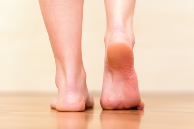
The history is very important in the setting of peroneal tendinosis. As noted above, these are overuse injuries. People with peroneal tendinosis typically have either tried a new exercise or have markedly increased their activities. Characteristic activities include marathon running or others which require repetitive use of the ankle. Patients will usually present with pain right around the back of the ankle. There is usually no history of a specific injury.
As discussed above, improper training or rapid increases in training and poor shoewear can lead to peroneal tendinosis. Also, patients who have a hindfoot varus posture may be more susceptible. This is because in those patients, the heel is slightly turned inwards which requires that the peroneal tendons work harder. Their main job is to evert or turn the ankle to the outside, which fights against the varus position. The harder the tendons work, the more likely they are to develop tendinosis.
There are two peroneal tendons that run along the back of the fibula (Figures 1 and 2). The first is called the peroneus brevis. The term “brevis” implies short. It is called this because it has a shorter muscle and starts lower in the leg. It then runs down around the back of the bone called the fibula on the outside of the leg and inserts (i.e. connects) to the fifth metatarsal. This is in the side of the foot. The peroneus longus takes its name because it has a longer course. It starts higher on the leg and runs all the way underneath the foot to insert or connect on the first metatarsal on the other side. Both tendons, however, share the major job of everting or turning the ankle to the outside. The tendons are held in a groove behind the back of the fibula and have a roof made of ligamentous-type tissue over the top of them called a “retinaculum.”